Keratoconus

Keratoconus

Keratoconus

Keratoconus

Keratoconus

Keratoconus

Keratoconus

Keratoconus
What Is Keratoconus?
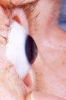
Keratoconus, often referred to as “KC,” is a non-inflammatory eye condition in which the typically round dome-shaped cornea progressively thins and weakens, causing the development of a cone-like bulge and optical irregularity of the cornea. This causes “static” in your vision and can result in significant visual impairment.
Symptoms
Keratoconus typically first appears in individuals who are in their late teens or early 20s, and may progress for 10 to 20 years and then slow or stabilize. Each eye may be affected differently. In the early stages of keratoconus, people might experience:
Slight blurring of vision
Distortion of vision
Increased sensitivity to light
The cornea is responsible for focusing most of the light that comes into the eye. Therefore, abnormalities of the cornea, such as keratoconus, can have a major impact on how an individual sees the world, making simple tasks such as driving a car or reading a book very difficult.
In keratoconus care, early detection is critical because of the condition’s progressive nature. Your optometrist plays a key role in the diagnosis of keratoconus because the sooner they identify the signs and symptoms, the sooner they can refer you for treatment that can slow or halt disease progression to help preserve your vision.
Keratoconus:
Can result in significant vision loss
May lead to corneal transplant in severe cases
Affects both males and females
Affects all ethnicities
10% of people with KC have affected relatives
People with Down syndrome are 20 times more likely to be affected
You can find more information about keratoconus at livingwithkeratoconus.com.
What Is iLink™ Corneal Cross-Linking?

The American Academy of Ophthalmology Corneal Ectasia Preferred Practice Pattern recommends prompt referral of patients who have been diagnosed with progressive keratoconus by their optometrist to a trusted ophthalmologist who can perform corneal cross -linking.3 iLink™ corneal cross-linking is a minimally invasive outpatient procedure that combines the use of ultraviolet light and specially formulated eye drops to stiffen and strengthen corneas that have been weakened by disease or refractive surgery. Cross-linking is considered the standard of care around the world for progressive keratoconus and corneal ectasia following refractive surgery.
Is iLink™ Covered by Insurance?
The medical necessity of iLink™ has become widely recognized. As a result, commercial insurance coverage for iLink™ is now over 95% in the United States for covered lives. Only iLink™ is approved by the FDA and is covered by insurance. Typically, non–FDA-approved procedures are not covered by insurance and have not been proven safe and effective.
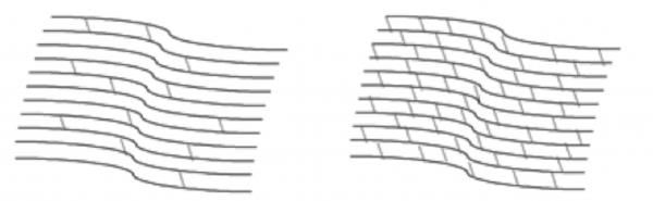
Corneal Cross-Linking
Creates new corneal collagen cross-links
Results in a shortening and thickening of the collagen fibrils
Leads to the stiffening of the cornea
Riboflavin
Under the conditions used for iLink™ corneal cross-linking, specially formulated pharmaceutical-strength riboflavin eye drops called Photrexa® (riboflavin 5’-phosphate ophthalmic solution) and Photrexa® Viscous (riboflavin 5’- phosphate in 20% dextran ophthalmic solution) help enable the cross-linking reaction.
Ultraviolet Light (UV)
iLink™ corneal cross-linking applies an artificial source of UV light from a machine called the KXL® System once the cornea has been soaked in the Photrexa® and Photrexa® Viscous eye drops. This process works to stiffen the cornea by increasing the number of molecular bonds, or cross-links, in the collagen.
Combining Riboflavin and UV Light
Using Photrexa® and Photrexa® Viscous riboflavin eye drops, combined with ultraviolet light from the KXL® system, the iLink™ procedure stiffens and strengthens the cornea to slow or halt progressive keratoconus.
Is Cross-Linking Right for Me?
Patients who have been diagnosed with progressive keratoconus or corneal ectasia following refractive surgery should be promptly referred to an ophthalmologist who can perform corneal cross-linking. Our practice partners with an ophthalmologist who performs iLink™ corneal cross-linking. This ensures that you can be confident that you are receiving the only FDA-approved cross-linking procedure. Speak with your doctor about the risks and benefits of iLink™ to determine if it may be right for you.
The goal of iLink™ is to slow or halt the progression of keratoconus to help preserve patients’ vision. Typically, within 1 or 2 months after an iLink™ procedure, patients will return to their optometrist for ongoing monitoring and overall vision care.
For more information about the iLink™ procedure for the treatment of keratoconus and corneal ectasia following refractive surgery, visit livingwithkeratoconus.com.
Why iLink™?
Not all corneal cross-linking procedures are the same. Only iLink™ is approved by the FDA for the treatment of progressive keratoconus and backed by robust clinical studies. Other unapproved cross-linking procedures have not been rigorously studied and are not supported by the same standards of clinical efficacy and safety. Additionally, non–FDA-approved cross-linking procedures may be associated with greater out-of-pocket costs for patients.
Photrexa® Products and KXL® System Photos
Using Photrexa® Viscous, Photrexa®, and the KXL® system, the iLink™ corneal cross-linking procedure from Glaukos is the only FDA-approved therapeutic treatment for patients with progressive keratoconus and corneal ectasia following refractive surgery.
INDICATIONS
Photrexa® Viscous (riboflavin 5’-phosphate in 20% dextran ophthalmic solution) and Photrexa® (riboflavin 5’- phosphate ophthalmic solution) are indicated for use with the KXL System in corneal collagen cross-linking for the treatment of progressive keratoconus and corneal ectasia following refractive surgery. Corneal collagen cross-linking should not be performed on pregnant women.
IMPORTANT SAFETY INFORMATION
Ulcerative keratitis can occur. Patients should be monitored for resolution of epithelial defects. The most common ocular adverse reaction was corneal opacity (haze). Other ocular side effects include punctate keratitis, corneal striae, dry eye, corneal epithelium defect, eye pain, light sensitivity, reduced visual acuity, and
blurred vision.
These are not all of the side effects of the corneal collagen cross-linking treatment. For more information, go to www.livingwithkeratoconus.com to obtain the FDA-approved product labeling.
You are encouraged to report all side effects to the FDA. Visit www.fda.gov/medwatch, or call 1-800-FDA-1088.
References:
1. Understanding KC. National Keratoconus Foundation. Accessed April 1, 2021. https://nkcf.org/understanding-kc.
2. Gelles JD. The Optometrist’s Role in Keratoconus Management. Advanced Ocular Care. April 2017.
3. Garcia-Ferrer FJ, Akpek EK, Amescua G, et al; American Academy of Ophthalmology Preferred Practice Pattern Cornea and External Disease Panel. Corneal Ectasia Preferred Practice Pattern®. Ophthalmology. 2019;126(1):P170-P215.
4. Gomes JAP, Tan D, Rapuano CJ, et al. Global consensus on keratoconus and ectatic diseases. Cornea.
2015;34(4):359-369.
5. Beshtawi IM, O’Donnell C, Radhakrishnan H. Biomechanical properties of corneal tissue after ultraviolet-A-riboflavin crosslinking. J Cataract Refract Surg. 2013;39(3):451-462

All Eye
Care Services

Keep
In Touch
Our Services
Contact Info
Hours of Operation
- Monday 8:00 AM - 6:00 PM
- Tuesday 8:00 AM - 6:00 PM
- Wednesday 8:00 AM - 6:00 PM
- Thursday 8:00 AM - 6:00 PM
- Friday 8:00 AM - 3:00 PM
- Saturday Closed
- Sunday Closed
© 2024 Premier Eye Care. All Rights Reserved. Accessibility Statement - Privacy Policy - Sitemap
Powered by:
